Whitepaper: The Next Dimension of Discovery: Advancing Spatial Biology in Three Dimensions
See how going 3D gives you a deeper understanding of your tissue
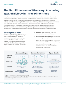
Transcriptomic analysis has traditionally relied on single-cell analysis or 2D thin sections. While informative, these methods give an incomplete picture of what’s going on in the tissue. Rare events can remain hidden, spatial distances and adjacencies can be misrepresented, and cell morphologies can be disrupted.
To overcome these limitations, 3D spatial biology methods in intact tissue helps scientists study cells, transcripts, proteins, and their interactions in a more native environment. With 3D spatial biology, you’ll get:
✔ A better representation of true biological conditions
✔ Increased cell density and cell-cell proximity measurements
✔ More data within the same spatial area
In this white paper, see how 3D spatial biology can help you uncover rare events, better represent spatial relationships, and visualize transcription and translation within the cell.

True 3D™ spatial transcriptomics, only on Pyxa.
Subscribe to the Stellaromics newsletter to stay at the forefront of 3D spatial biology.
- (857) 675-9157
- info@stellaromics.com
- 321 Harrison Ave, 5th Floor, Boston, MA 02118