Analysis in Every Dimension
Discover the only technology that creates fully annotated 3D maps of gene expression within thick tissue samples.
Tissue is not paper thin.
It's time to stop studying
it that way.
Spatial biology is a significantly disruptive technology, but so far, we’ve only scratched the surface – literally.
Tissue is not only composed of complex and diverse individual cells, but of layers of those cells all interacting in three dimensions with profound biological relevance.
In order to truly understand biology, our analysis must be both broad and deep, encompassing single cell analysis across multiple layers of tissue.
Go beyond the surface.
Capture truly multidimensional cellular dynamics.
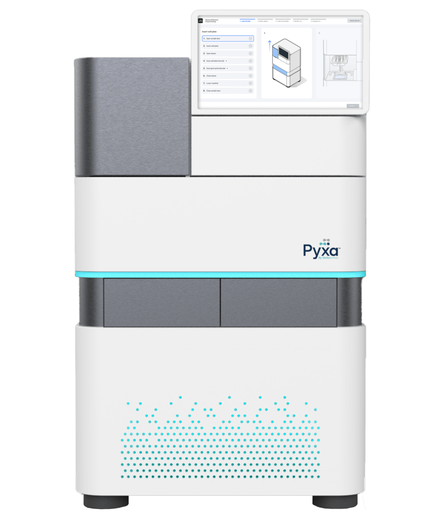
Pyxa is designed for 3D spatial multiomics
Sample preparation, automated volumetric confocal imaging, data analysis and visualization all in one integrated platform.
- Process up to 12 wells simultaneously
- For a multitude of sample types
- Automated image processing
- Advanced data visualization


“We are thrilled to be among the first to utilize the groundbreaking Pyxa platform. A key advantage of Pyxa over other spatial transcriptomic technologies is its ability to analyze much larger tissue volumes per experiment. By enabling dense reconstructions of intact neural circuits in 3D, Pyxa will significantly advance neuroscience research, particularly for our laboratory's goal of reconstructing cell type-specific synaptic connectivity relationships in high-throughput by tracking the synaptic spread of viruses using RNA barcoding.”
Arpy Saunders
Assistant Professor at the Vollum Institute of OHSU

“As an early user of the Pyxa platform, I’ve been impressed by its ability to deliver a comprehensive 3D perspective on biological systems. The platform is fundamentally useful, as tissue analysis is inherently three-dimensional. We’re excited to continue partnering with Stellaromics to push the boundaries of scientific research.”
Gordon Wang, PhD
Clinical Associate Professor, Stanford University

“The transition from 2D to 3D spatial omics is transformative for neuroscience. While 2D methods provide molecular profiles, they miss critical long-range cellular interactions. We can now visualize these connections at an unprecedented scale, enabling high-throughput analysis of genetic perturbations across complex tissues. Combining CRISPR gene editing with high-resolution spatial analysis allows us to uncover new insights into brain development and disease progression in ways we never could before.”
Xin Jin MD, PhD
Associate Professor at Scripps Research

“I’m incredibly excited about the potential of Stellaromics’ 3D spatial
multiomics platform for advancing cancer research. Its ability to analyze
thick tissue sections in 3D offers unprecedented insights into tumor heterogeneity
and the tumor microenvironment, including the progression of pre-malignant
pancreatic cysts and healthy liver tissue to tumor. Visualizing complex cellular
interactions in 3D is crucial for our work on colorectal liver metastasis and
hepatocellular carcinoma. I’m particularly eager to explore the platform’s
capacity to detect rare cell populations, such as infiltrating immune cells. 3D
spatial represents a transformative step toward building comprehensive disease
atlases, ultimately improving patient care through more precise diagnostics and
targeted therapies.”
Nigel Jamieson, MD
Professor of Surgery, University of Glasgow
3D spatial omics answers biological questions that no other technology can.
Meet with our team to discuss applications for Pyxa in your research, and how to start a services project or purchase an instrument.