
Pyxa®: True 3D™ spatial multi-omics in thick tissue
Sample preparation, automated imaging, and 3D data analysis all in one platform.
Now commercially available.
Capture the full complexity of diverse cellular landscapes with True 3D™ spatial multiomics on Pyxa
Pyxa is the first platform for high-plex spatial multi-omic analysis of thick tissue sections in 3D. Pyxa is an automated, high-plex, confocal spatial platform designed to deliver fully annotated, single-cell maps of 3D gene expression in thick tissue.
The end-to-end system was intentionally designed to automate the STARmap™ protocol for ease of use in labs and core facilities.
An end-to-end system for high-plex 3D spatial multi-omics
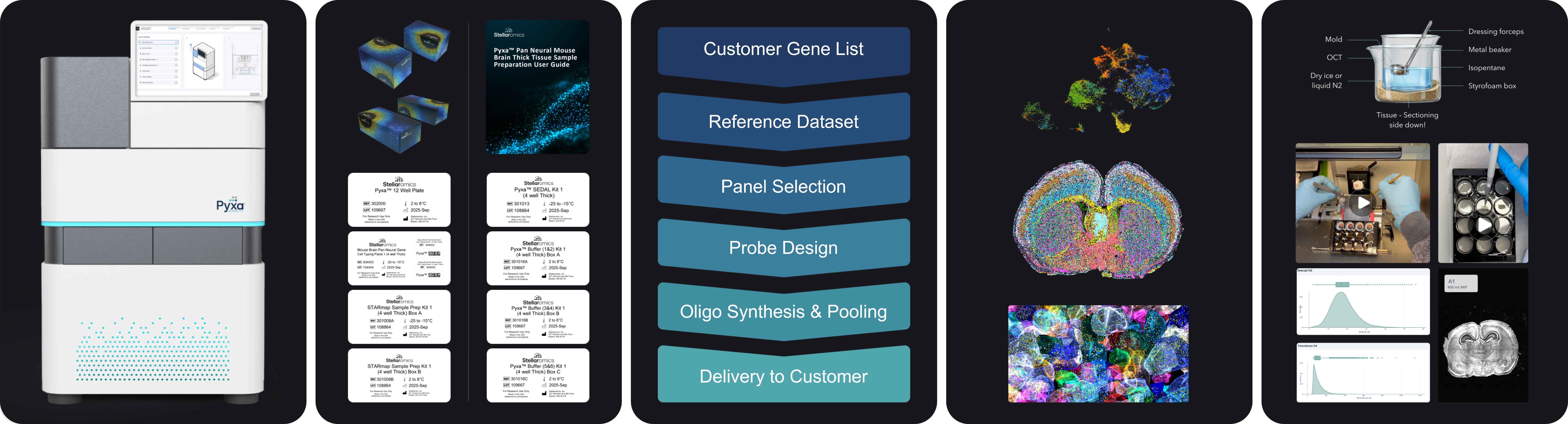
3D spatial biology on Pyxa starts with thick, intact tissue sections (up to 100 µm) – up to 20 times thicker than the 5-10 µm tissue sections allowed by other platforms.
Off-the-shelf or custom gene panels, combined with kits for sample preparation, make it easy to get started. Sample preparation is in a 12-well microplate format. We currently offer sample preparation kits for 100 micron thick tissue, and an off-the-shelf panel for mouse brain. For other tissue types, customers can provide Stellaromics with a gene list for custom panel design and synthesis.
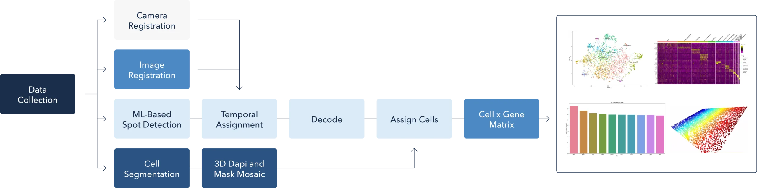
Once a sample has been processed, 3D spatial omics data can be explored using the PyxaStudio data visualization software included on the instrument workstation. Data output files are also compatible with numerous tools for single cell and spatial transcriptomic data analysis.
Pyxa is built on STARmap™ chemistry
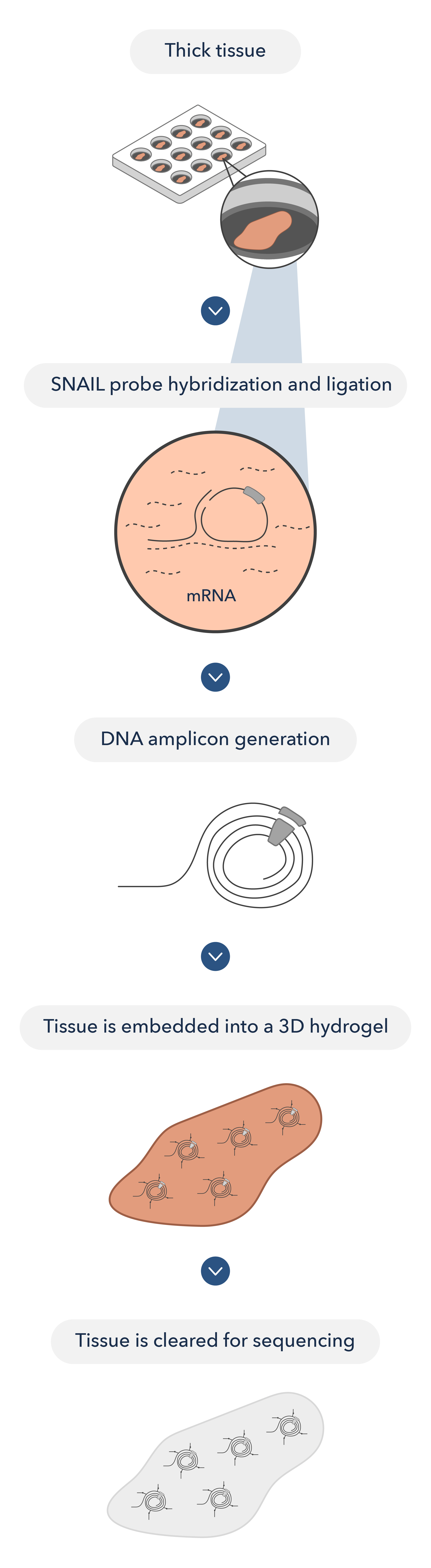
STARmap chemistry leverages specific amplification of nucleic acids via intramolecular ligation using paired primer and padlock probes (SNAIL probes) to convert target RNA molecules into DNA amplicons with gene-unique barcodes. This enables highly multiplexed RNA detection in tissue hydrogels by multiple rounds of sequencing by ligation.
Flexible chemistry allows targeting of both short and long RNA sequences, whether endogenous or exogenous.
3D sequencing by ligation with error-reduction
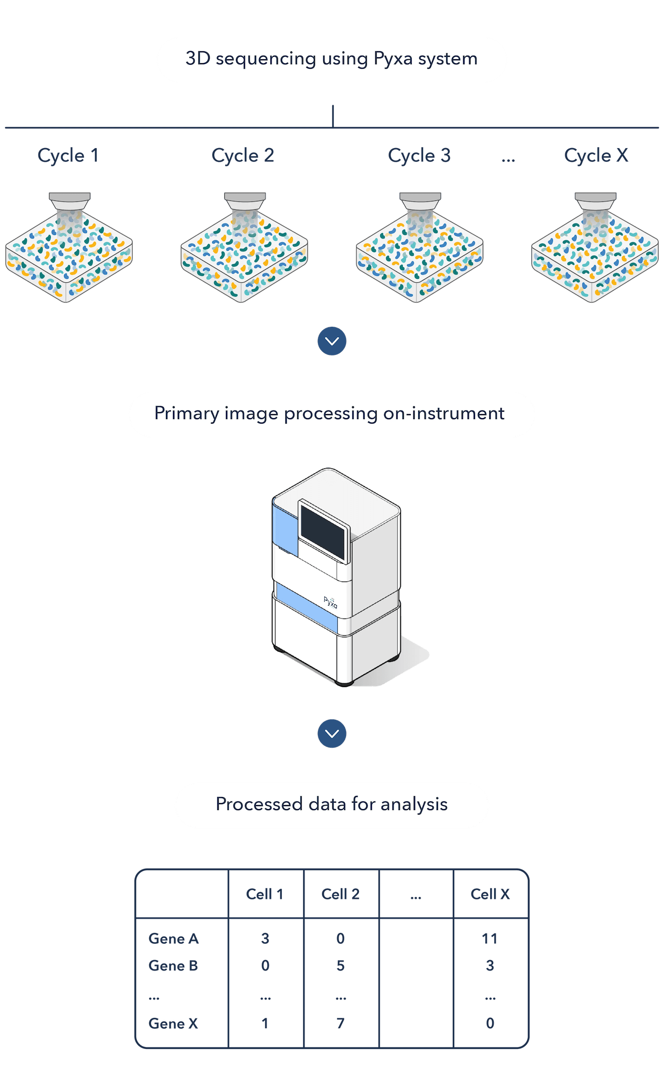
Sequencing with error-reduction by dynamic annealing and ligation (SEDAL) uses reading probes to decode bases, and fluorescence probes to decode sequence information into fluorescence signals.
Decoding probes only transiently bind to the target DNA and ligate to form a stable product for imaging only when a perfect match occurs, which eliminates error accumulation.
Assays available on the Pyxa platform
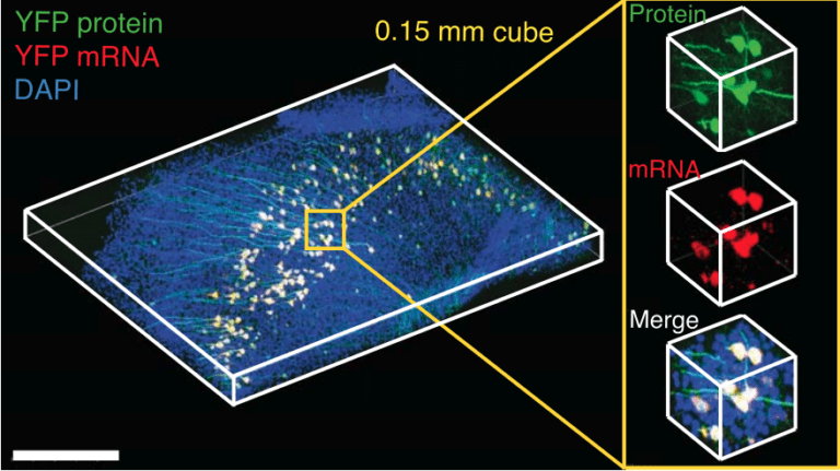
STARmap for spatial transcriptomics
Challenge: Retrieving high-content gene-expression information while retaining 3D spatial localization at subcellular resolution
Innovation: 3D intact-tissue RNA sequencing, which integrates proprietary hydrogel-tissue chemistry, targeted signal amplification, and in situ sequencing.
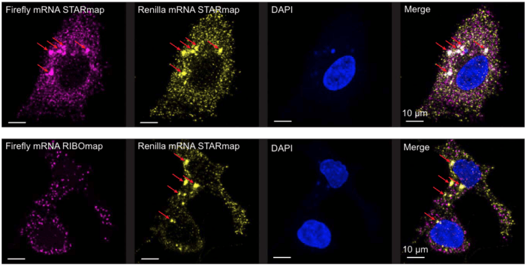
RIBOmap for spatial translatomics
Challenge: RNA levels and protein levels often do not match up. Profiling proteins is costly and difficult to scale to large numbers.
Innovation: RIBOmap measures only translated RNA, serving as a practical and scalable approximation of protein expression, with 3D localization.
Advanced visualization and analysis of 3D spatial multi-omic data
From 3D Images to Spatial Insights using the PyxaStudio Data Visualization Software
PyxaStudio is our dedicated visualization tool for navigating 3D tissue architecture – from broad cellular organization down to subcellular transcript localization – all within the native volumetric context of the tissue. Key features of PyxaStudio include:
- Visualize multiple cell layers in 3D.
- Explore the spatial distribution of cells and transcripts throughout the tissue.
- Investigate subcellular transcript localization.
- Subselect cells within UMAP or spatial view.
- Bookmark cells and cell neighborhoods of interest and share them with collaborators.
Data output files are compatible with numerous open-source tools for single cell and spatial transcriptomic data analysis
- Cell-by-gene matrix
- 3D gene expression patterns in intact tissue, including per-transcript XYZ coordinates and cell assignments
- Cell volumes and centroid positions in XYZ
Frequently Asked Questions
Most spatial technologies are capable of imaging 5-10 µm thin tissue slices. But cells and tissues are three-dimensional, and these platforms destroy this biology instead of capturing it.
Pyxa images intact, 100 µm thick tissue sections. Instead of slicing through cells and performing computationally-intensive reconstruction of serial sections, it’s possible to capture full cell volumes and intact tissue morphology on Pyxa.
Pyxa can image tissue sections up to 100 microns thick. We currently offer sample preparation kits for 20 micron and 100 micron thick tissue sections.
STARmap on Pyxa allows researchers to image intact tissue sections up to 100 microns thick- up to 20 times thicker than is possible using 2D spatial transcriptomics platforms. This permits the visualization of multiple intact cellular layers, capturing rare cells, edit events, and complex structures that are obscured when sectioning tissue into thin, 5-10 micron slices.
2D cell segmentation fundamentally misrepresents cell biology because it can only capture cross-sections of each cell. This is because cells are larger than the 5-10 µm thickness that other spatial platforms can image.
3D cell segmentation captures the full volume of each cell. This is critical for analyzing complete information about each cell, deconvoluting adjacent cell types, and visualizing cell shapes, process extensions, and true neighborhood relationships.
We currently offer commercial products for detecting RNA transcripts on Pyxa. We anticipate launching products for protein co-detection on Pyxa in late 2026. You can contact our team now to register your interest!
Yes, tissue clearing is required as part of the STARmap protocol. Embedding the tissue in hydrogel to fix amplicons in 3D space, followed by tissue clearing to remove proteins and lipids from the sample, is what ensures the accurate detection of RNA transcripts in thick tissue sections in 3D space on Pyxa.
Yes – please contact us for access to 3D spatial transcriptomic data from Pyxa.