
3D Spatial Multi-omics for Neuroscience

Pyxa maps RNA, translation and protein in 100 μm brain tissue – so cells, circuits and cortical layers stay intact.

The brain isn't flat. Your spatial data shouldn't be either.

Neurons, astrocytes, vasculature and cortical layers extend across many cell layers. When spatial transcriptomics is confined to a 5–10 μm section, large cells are sliced through, projections are truncated, and the three-dimensional organization that defines brain tissue is lost before the analysis begins.

Enter Pyxa.

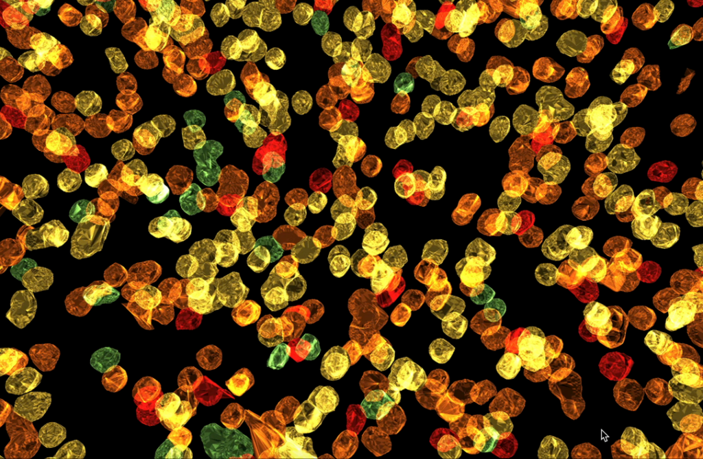
Pyxa is purpose-built for profiling intact tissue. It handles 100 μm sections — up to 20x thicker than conventional spatial platforms — with confocal imaging at subcellular resolution. Complete cells, preserved neuronal projections, the architecture of cortical layers, and the cellular neighborhoods around amyloid beta plaques and tau tangles are captured in their native context.

Pyxa Features

Flexible Gene Panel Design for Neuroscience Research on Pyxa

Off-the-shelf

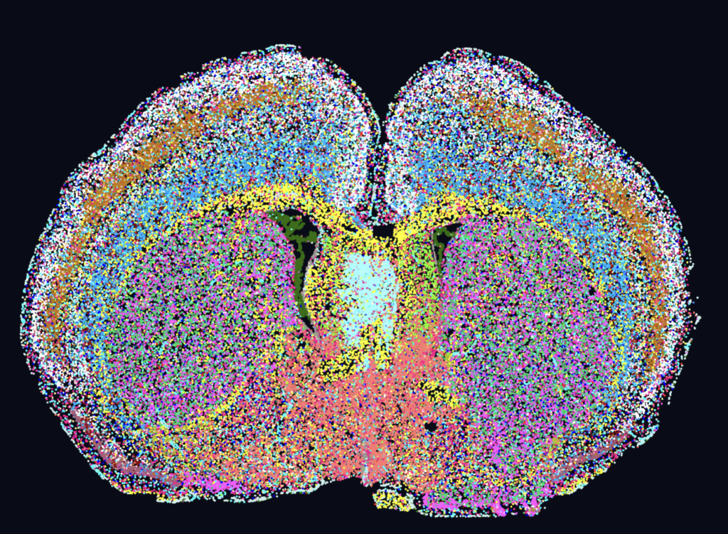
237-gene core panel for mouse brain, designed to subset key cell populations in the brain.

Customizable

By expanding the mouse brain core panel with custom genes, users can identify rare cell populations and understand the spatial distribution of cell types in the mouse brain.

Fully Custom

Researchers can design fully custom gene panels for human or mouse brain research.

Pyxa is Accelerating Discovery in Neuroscience and Neurodegeneration Research

Neurodegeneration: Cellular neighborhoods around amyloid-β plaques, captured whole

Researchers at the University of Virginia combined STARmap with protein co-detection to map the cells surrounding amyloid-β plaques in a mouse model of Alzheimer’s disease — resolving which cell types and transcriptional states cluster around disease pathology and how those neighborhoods change as disease progresses. Because every plaque is captured in 3D with its full complement of neighboring cells, true plaque-associated states are counted, not inferred from a slice.

Addiction & Behavior: Neuronal aggregates in the nucleus accumbens, visible only in 3D

Researchers from the Ichan School of Medicine at Mount Sinai used 3D spatial transcriptomics on Pyxa to identify isolated versus aggregated neurons in the nucleus accumbens, a key brain region in addiction. These clusters of neurons could not be visualized by 2D omics methods and could only be identified by visualizing the brain in 3D.

Neural Circuits: Viral tracing meets cell-type identity in intact tissue

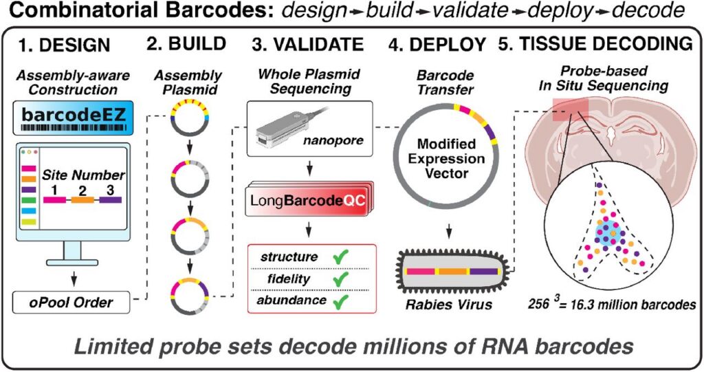
Viral tools reveal cell-cell connectivity; Pyxa localizes that readout in intact tissue. Labeled neurons and their projections can be mapped alongside transcriptomic cell identity across a 100 μm volume, linking circuit membership to molecular state.  The combinatorial barcode design and assembly framework developed by the Saunders Lab is well-suited for designing new types of barcoding schemes to meet the needs of existing and future probe-based genomics technologies.

Hear from neuroscientists who have used Pyxa

Pyxa 3D has been an outstanding tool for our work. The ability to visualize spatial separations in three dimensions has been a game-changer. We've applied it across multiple brain regions and consistently had a positive experience, and the platform has performed equally well with our custom panels. The PyxaStudio visualization software has also been excellent for viewing and exploring the data.

Lauren Wills

Assistant Professor of Neuroscience, Icahn School of Medicine at Mount Sinai

We are thrilled to be among the first to utilize the groundbreaking Pyxa platform for highly multiplexed experimentation in the brain. A key advantage of Pyxa over other spatial transcriptomic technologies is its ability to analyze large brain tissue volumes, enabling more features of brain cells - such as cellular morphology - to be tracked alongside molecular properties. Linked analysis of multiple thick tissue sections enables dense reconstructions of intact neural circuits in 3D, opening up new experimental opportunities, particularly for our laboratory's goal of reconstructing cell type-specific synaptic connectivity relationships in high-throughput by tracking the synaptic spread of individual viruses using RNA barcoding.

Arpiar Saunders

Assistant Scientist, Vollum Institute

3D spatial transcriptomics is transformative for our Alzheimer’s disease research because it allows us to investigate pathology within its native cellular and anatomical context. In our APP/Tau double-transgenic model, we can map how amyloid and tau pathology are associated with distinct neuronal and glial subpopulations across different brain regions. Rather than viewing these changes as isolated molecular signals, 3D spatial data allow us to resolve how pathology, cell states, and regional vulnerability are interconnected. This level of spatial resolution is critical for understanding the cellular heterogeneity and disease mechanisms that conventional 2D approaches cannot fully capture.

Lulu Jiang

Assistant Professor, University of Virginia School of Medicine
Request a 3D demo

Talk to our team about how 3D spatial multi-omics can answer your neuroscience questions — and what your first 100 μm dataset could look like.

Frequently Asked Questions

What are the sample requirements for brain tissue on Pyxa?

Pyxa supports fresh frozen brain tissue from mouse, human, and NHP.

For information on the latest tissue fixation and species compatibility, please contact us.

Pyxa supports tissue sections up to 100 µm thick, and kits are designed to support 100 µm tissue. It is possible to use thinner sections, such as 20 µm or 50 µm, on the instrument.

Yes, you can add up to 20 custom genes to the standard mouse brain panel. You can also design a fully-custom panel, which has a 6-8 week lead time for panel design.

Stellaromics offers PyxaStudio™, a bespoke data visualization software designed to explore 3D spatial data. Data files are compatible with common secondary analysis pipelines in R and Python including Seurat and Scanpy.